RIGHT SIDED CYSTIC BREAST MASS
Formerly Published in Vol. 6 No. 1 Jan. - Jun., 2021
DOI:
https://doi.org/10.83120/pmj.vol3no1.21Keywords:
Breast cyst, mammography, Doppler imaging, radiological assessment, breast pathologyAbstract
Background: Cystic breast mass, characterized by fluid-filled sac within breast tissue, is a frequent occurrence in clinical practice. This case report delves into the radiological diagnosis ofa cystic breast mass emphasizing the necessity for a thorough radiological evaluation. Cystic breast lesions are identified using the following imaging modalities like mammography, ultrasonography, and magnetic resonance (MR) imaging.It is important to note thatboth benign simple cyst, complex cystic and solid masses needs to be biopsied. Breast Imaging Reporting and Data System (BI-RADS)[1] is used in the characterization of breast lesions. This case report explores the clinical presentation radiological features, and diagnostic considerations, underscoring the crucial role of accurate imaging techniques in managing cystic breast masses. The findings in this case report contribute to a better understanding of breast pathology and highlights the importance of precise diagnostic approaches in clinical decision-making.This will go a long way in early detection of breast lesions and better management and outcome of patients.
Methods: A 25-year-old female with complaints of right breast pain and discomfort with palpable mass underwent a thorough clinical examination. Radiological investigations, including mammography,ultrasound of the breast and MRI (magnetic resonance imaging) were conducted to visualize and characterize the cystic masses. The imaging studies aimed to assess the size, location, and internal characteristics of the cystic mass, guiding subsequent diagnostic and management steps.
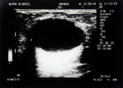
Results: Ultrasound of the breast revealed an anechoic mass with thick wall located in the right upper outer quadrant middle ring of the right breast measuring 51.2mm x 24.3mm in its dimensions, the lesion is wider than taller with no associated architectural distortion of the breast and no axillary lymphadenopathy. The mass displayed well-defined borders with no signs of micro calcifications. Ultrasound imaging also provided additional insights, confirming the cystic nature of the mass with posterior acoustic enhancement and excluding solid components, Doppler interrogation also revealed absence of blood flow showing that it is a non-vascular lesion.Mammography revealed a well-defined homogenous opacity in the right breast while MRI findings revealed that the lesion is of high signal intensity on T2weighted images and low signal intensity on fat suppressed T1 weighted images. The absence of vascularity on contrast enhanced T1 weighted images further supported the benign nature of the cystic mass. Histological findings after the excision of the cyst revealed benign features. A final (BI-RADS) assessment (BI-RADS) 2 was made.
Conclusion: The radiological evaluation of the cystic mass with histology highlighted the significance of accurate imaging and histology in distinguishing benign breast lesions from malignant lesions Clinical correlation and follow-up imaging is essential in ensuring appropriate management of the patient.
Downloads
References
1. Mendelson EB, Baum JK, Berg WA, et al. 1st edition American College of Radiology; Reston (VA): 2003. Breast imaging reporting and data system, BI-RADS: ultrasound. [Google Scholar] [Ref list]
2. W.A. Marshall, J.M. Tanner Variations in pattern of pubertal changes in girls Arch Dis Child, 44 (235) (1969), pp. 291-303, 10.1136/adc.44.235.291
3. E.J. Lee, Y.W. Chang, J.H. Oh, J. Hwang, S.S. Hong, H.J. Kim Breast lesions in children and adolescents: diagnosis and management Korean J Radiol, 19 (5) (2018), pp. 978-991, 10.3348/kjr.2018.19.5.978
4. E. Mareti, A. Vatopoulou, G.A. Spyropoulou, A. Papanastasiou, G.C. Pratilas, A. Liberis, et al. Breast disorders in adolescence: a review of the literature Breast Care, 16 (2) (2021), pp. 149-155, 10.1159/000511924
5. R.J. Brenner, M.E. Bein, D.A. Sarti, A.L. Vinstein Spontaneous regression of interval benign cysts of the breast Radiology, 193 (2) (1994), pp. 365-368
6. W.A. Berg, A.G. Sechtin, H. Marques, Z. ZhangCystic breast masses and the ACRIN 6666 experience Radiol Clin, 48 (5) (2010), pp. 931-987
7. P.C. Stomper, B.J. Van Voorhis, V.A. Ravnikar, J.E. Meyer Mammographic changes associated with postmenopausal hormone replacement therapy: a longitudinal study Radiology, 174 (2) (1990), pp. 487-490
8. Berg WA, Blume JD, Cormack JB, et al. Combined screening with ultrasound and mammography vs mammography alone in women at elevated risk of breast cancer. JAMA. 2008;299(18):215163. [PMC free article] [PubMed] [Google Scholar]
9. Venta LA, Kim JP, Pelloski CE, et al. Management of complex breast cysts. AJR Am J Roentgenol. 1999;173(5):1331–6. [PubMed] [Google Scholar]
10. A. Jakubowska, M. Grajewska-Ferens, M. Brzewski Breast cysts in adolescents - diagnostics, monitoring, treatment Pol J Radiol, 76 (1) (2011), pp. 20-24
Downloads
Published
Issue
Section
License
Published articles are distributed under the Creative Commons Attribution 4.0 International (CC BY 4.0) licence, unless otherwise stated.